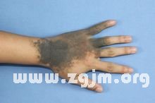
Melanin cell nevus, melanin nevus
Melanin cell nevus, melanin nevus

Melanin mole
Melanin nevus (melanocytic nevi) is composed of a group of benign melanocytes, gathered at the border, epidermal and dermal melanin cells may be distributed at the bottom of the reticular dermis (the lower reticular diemis), connective tissue between the beam (between collagen bundles), around the skin of the other affiliated organs such as sweat glands and hair follicles, blood vessels, nerves, etc., occasionally to the subcutaneous fat.
Its appearance looks may be flat, convex, verrucous, granular, or other shapes, colors may be brown, black and blue.Melanin nevus have two kinds of produced nature and nurture.
Congenital nevus at birth or the neonatal period is mostly, acquired nevus is after six months, until old age is likely to be new.
Acquired nevus size zero point one to zero point six cm, usually on the pathology can be divided into three kinds, joint nevus (limited nevus cells in the epidermis, dermis border area, within the epidermal nevus), compound nevus (nevus cells not only distributed in the epidermis layer, down into the dermis), intradermal nevus (nevus cells in the dermis completely inside).Clinical appearance of moles also related to the pathological classification, joint birthmark rendering brown to black flat spots, not bumps on the skin surface, usually brown ridges of compound nevus papule or nodules, intradermal nevus is bigger, more bumps, a light brown or flesh-colored nodule, common people, which is "mole" meat.
In 1% ~ 2% of all births, congenital nevus melanin can be found, but the incidence of giant congenital nevus melanin is less than one over twenty thousand (statistics) abroad.Clinically can be in accordance with the size of the melanin mole divided it into three types:
Small mole melanin: size is less than two centimeters, preference distribution in the lower body, upper back, shoulders, chest and proximal limb.
Intermediate melanin mole: size between 2 ~ 20 cm.
Giant nevus melanin: the distribution of size greater than 20 cm, with the second half of the trunk is given priority to, also somebody won't, in the head or other limbs may cover large skin of the body.Are usually dark, and there are some hair covering, outside the main body is also dotted with satellite lesions.
Most of the congenital giant nevus melanin is benign, but congenital giant nevus melanin are usually more complicated than acquired huge melanin nevus.According to the growth of its type and can be divided into three types:
Compound or subcutaneous nevus (compound or intradermal nervus) :The most commonly occurs.
Nerves nevus (neural nervus) :Neural tube or nerve tumors can occur in the structure, looks like a nerve fibroma.
Blue nevus (blue nervus) :The most rare.
Melanin nevus in clinic must do distinguish, and melanoma melanoma is the highest mortality rates of skin cancer, about two-thirds of all patients with skin cancer deaths.According to the west is about 20 ~ 50% of melanoma and nevus.So tell whether moles merger melanoma is very important.General according to the ABCDE will distinguish, A is asymmetric, Asymmetry is asymmetrical lesions up and down, left and right sides (imagine lesions can control or up and down like origami folded).B is Border Irregularity irregular edge, edge is not form a circular arc form, and appear jagged gaps;C Color Variability Color is uneven, some parts of the pigment, some parts of the shallow pigment;D is the Diameter > 6 mm, the Diameter of the lesion is greater than zero point six cm;E is Elevation or Enlargement, namely surface become bumps or lesion size increased.For any medically suspected lesions may have malignant lesions, have to accept a biopsy and pathology assay.Fortunately, in the Asian race, melanoma with nevus, much less than caucasians.
Causes and prevention
Ultraviolet (uv) is a major source of melanin formation, at the same time of whitening care in daily life to prevent bask in, can well restrain melanin formation.Because at the time of whitening care, would have been generated melanin slowly elimination, and sunscreen to help curb new skin melanin formation.Through good sunscreen to protect the base layer of skin and the dermis from uv rays, does not stimulate the activity of tyrosinase, namely can inhibit the generation of melanin.New and old cutin alternating slow also is one of the causes of the formation of melanin, so regularly purify is corneous, it will be very helpful for whitening skin, adding acid, now a lot of beautiful white product is helpful, peel off aged horniness, eliminate melanin.
Through diet can also improve color of skin.Such as a large number of drinking water, eat more vegetables and fruits at ordinary times, enhance antioxidant food intake, eat foods rich in vitamin C.Chemical experiments show that the melanin formation of a series of oxidation reaction, but when adding vitamin C, can block the formation of melanin.Therefore, eat more foods rich in vitamin C, such as zizyphus jujube and fresh jujube, tomato, pear, citrus, fresh green leafy vegetables, etc.
In medicine melanin nevus belongs to benign tumors, generally do not need treatment.Want to remove melanin mole patients, the best choice to treat skin specialists and have safeguard relatively.Commonly used treatment, surgery, laser therapy, such as radio knife resection.
Melanin nevus easily confused with what disease?
1. The border benign nevus
Seen microscopically for benign nevus cells is not the opposite sex only grow in the leather in the inflammatory reaction is not obvious
2. Childhood sexual melanoma
inThe childFace of slow growth in circular nodules microscopically, the cellular pleomorphism with nuclear fission tumor cells to nor formation on the surface of skin infiltration and tumors had ulcer
3. Blue nevus cell sex
Occurs in hip tail sacral pale blue waist irregular nodular surface smooth and microscopically deep black of the dendritic cells and collection into large prismatic cells island is a fission or necrosis area should take into account the possibility of malignant change
4. Basal cell carcinoma
Is epithelial malignant tumor from the base layer of skin to deep infiltrating nests for the layer of columnar or cuboidal cells around the cancer cell staining deep procession is arranged within the cancer cells may contain melanin
5. Sclerosing hemangioma
Excessive epidermal keratinization leather emulsion proliferation of expansion of the capillaries is often down skin tu around seemingly hematoma within the sample
6. The elderly nevus
In the old surface show verrucous nevus is excessive diversification grain layer part of stratum spinosum thickening or atrophy hypertrophy grassroots complete can also increase the pigment appearance of dermal papillary hyperplasia is a papilloma hyperplasia
7. Seborrheic keratosis
Was also papilloma focal hyperplasia skin grain boundaries clear cornification incomplete layer thickening thinning or even disappeared after first hyperplasia of epidermis cells can have little or more of the pigment melanin
8. The hematoma under the nail bed
There are many corresponding injury history microscopically for dry cells can have epithelial hyperplasia of fibroblasts
- The last:Moles phobia
- Next up:Note there are those who after dot mole